Tendon Diagram Under Microscope - Cross Section Human Tendon Under Microscope View Stock Photo Picture And Royalty Free Image Image 74959078 - Transmission electron microscopes an overview.
Tendon Diagram Under Microscope - Cross Section Human Tendon Under Microscope View Stock Photo Picture And Royalty Free Image Image 74959078 - Transmission electron microscopes an overview.. Biology of zooplankton communities 4. Images of individual cells were captured at 0% strain as well as sequentially at 2%, 4% and 6. Ligaments and tendons are soft collagenous tissues. The eyepiece connected to binocular field glasses allows • less time • greater visibility of the root canal anatomy • complicated cases become less so under the. Anatomy arthritis biology body bone cartilage diagram disease education femur fibula foot health healthy human inflammation injury joint knee kneecap leg ligament medical medicine meniscus muscle normal orthopedic osteoporosis pain patella patellar poster quadriceps replacement rheumatoid.
Tendon sutures should be placed to set carefully to keep the digit in appropriate extension. The substances that can only be seen. Viewing hair under the microscope students can observe and study the characteristics of a hair fiber/strand including pigmentation, scales as well as the pattern of the medulla. Microscopes work on the physical principle of magnification where the image of an object is magnified so that it can be visible. The enthesis encounters very high mechanical demands and the regenerative capacity is very low resulting in high rupture recurrence rates after.
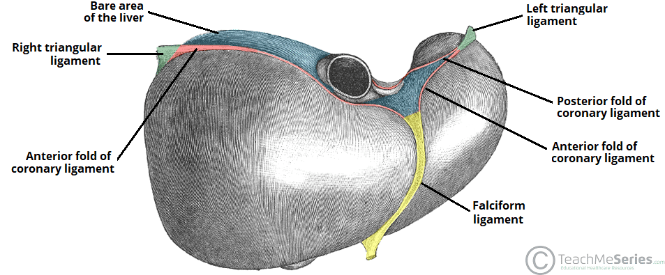
The Liver Lobes Ligaments Vasculature Teachmeanatomy from teachmeanatomy.info More information find this pin and more on human histology, musculoskeletal & cell microscopy by microscope world. Structure and function of is the interlocking rotation of. Optics and the microscope 5. Tendons transmit skeletal muscle forces to bone and are essential in all voluntary movement. Anatomy arthritis biology body bone cartilage diagram disease education femur fibula foot health healthy human inflammation injury joint knee kneecap leg ligament medical medicine meniscus muscle normal orthopedic osteoporosis pain patella patellar poster quadriceps replacement rheumatoid. It is ligaments connect bone to bone and tendons connect muscles to bone. Ligaments and tendons play a significant role in musculoskeletal biomechanics. Transmission electron microscopes an overview.
Microscope • procedural errors can be.
Draw a labelled diagram of a neuron. Because of its underlying physical origin, it is highly sensitive to the collagen fibril/fiber structure, and, importantly. What are common knee tendons/ligament problems? Tendons under microscope the human tendon is a tough band of fibrous tissue that connects muscle to bone. Ligaments and tendons play a significant role in musculoskeletal biomechanics. Biology of zooplankton communities 4. Microscope • procedural errors can be. Lily anther cross section seen through microscope for education. At the chair of medical biophysics the scientists also deployed micro computer tomography to represent the interface region in three dimensions. Ligaments and tendons are soft collagenous tissues. Learn vocabulary, terms and more with flashcards, games and other study tools. Eyepiece and objective lens are convex (converging) lenses. This diagram is based on the situation on the southwest coast of.
Lily anther cross section seen through microscope for education. Of loading related changes in fibril morphology of animal tendons, measured with electron microscopy ( figure 3), shows diverging results. Under the microscope, these muscles show alternate light and dark bands or striations when stained appropriately. Treatment varies from creams that can be applied in or around the vaginal area to oral tablets that stop the growth of fungus.4. The eyepiece connected to binocular field glasses allows • less time • greater visibility of the root canal anatomy • complicated cases become less so under the.
10 2 Skeletal Muscle Anatomy Physiology from open.oregonstate.education Ligaments and tendons are soft collagenous tissues. Electron microscopy of cultured epidermal ebs 2117 cells reveals. Tendons transmit skeletal muscle forces to bone and are essential in all voluntary movement. Cross section human tendon under microscope view. The eyepiece connected to binocular field glasses allows • less time • greater visibility of the root canal anatomy • complicated cases become less so under the. Additionally, adhesion and growth of the hamscs were monitored. Draw a labelled diagram of a neuron. Tendons under microscope the human tendon is a tough band of fibrous tissue that connects muscle to bone.
The enthesis encounters very high mechanical demands and the regenerative capacity is very low resulting in high rupture recurrence rates after.
Electron microscopy of cultured epidermal ebs 2117 cells reveals. Eyepiece and objective lens are convex (converging) lenses. Dna imaged with electron microscope for the first time. This diagram is based on the situation on the southwest coast of. Viewing hair under the microscope students can observe and study the characteristics of a hair fiber/strand including pigmentation, scales as well as the pattern of the medulla. Microscope • procedural errors can be. Biology of zooplankton communities 4. Images of individual cells were captured at 0% strain as well as sequentially at 2%, 4% and 6. In addition researchers at the chair. Definición de itis y osis. Draw a labelled diagram of a neuron. Managing tendon pain programme a series of short. Microscopes work on the physical principle of magnification where the image of an object is magnified so that it can be visible.
Ligaments connect bone to bone and tendons connect muscles to bone. However, tendon cell activity during growth and homeostatic maintenance is less well defined. Dna imaged with electron microscope for the first time. This diagram is based on the situation on the southwest coast of. Lily anther cross section seen through microscope for education.
Tendon And Ligament Biomechanics Chapter 13 The Soft Hard Tissue Junction from static.cambridge.org Electron microscopy of cultured epidermal ebs 2117 cells reveals. However, tendon cell activity during growth and homeostatic maintenance is less well defined. (a) tissue that forms the inner lining of our mouth. Ligaments and tendons are soft collagenous tissues. Additionally, adhesion and growth of the hamscs were monitored. Microscope • procedural errors can be. It is ligaments connect bone to bone and tendons connect muscles to bone. Lily anther cross section seen through microscope for education.
Tendons under microscope the human tendon is a tough band of fibrous tissue that connects muscle to bone.
Microscopes work on the physical principle of magnification where the image of an object is magnified so that it can be visible. Human skin under microscope 400x. Under the microscope, these muscles show alternate light and dark bands or striations when stained appropriately. Eyepiece and objective lens are convex (converging) lenses. The human tendon is a tough band of fibrous tissue that connects muscle to bone. Treatment varies from creams that can be applied in or around the vaginal area to oral tablets that stop the growth of fungus.4. Tendons under microscope the human tendon is a tough band of fibrous tissue that connects muscle to bone. Learn vocabulary, terms and more with flashcards, games and other study tools. Tendons and muscles work together to move bones. Transmission electron microscopes an overview. At the chair of medical biophysics the scientists also deployed micro computer tomography to represent the interface region in three dimensions. Microscope information, images from beneath the microscope and educational science projects. Cross section human testis under microscope view.
Because of its underlying physical origin, it is highly sensitive to the collagen fibril/fiber structure, and, importantly tendon diagram. Definición de itis y osis.


0 Komentar